3D Cone beam Imaging Adamsville
See Your Oral Structures in Amazing Detail with Advanced 3D Conebeam Imaging in Adamsville, TN
Get the most accurate diagnosis and precise treatment planning possible with revolutionary 3D imaging technology that reveals hidden problems and allows for safer, more predictable dental procedures with superior outcomes.
Revolutionary Benefits of 3D Conebeam Imaging
- Complete 3D Visualization: See teeth, bones, nerves, and tissues from every angle with incredible detail
- Precise Treatment Planning: Plan implants, extractions, and surgery with millimeter accuracy before treatment begins
- Enhanced Safety: Locate vital structures like nerves and sinuses to avoid complications during procedures
- Faster Diagnosis: Detect problems that traditional 2D X-rays might miss or require multiple images to reveal
- Reduced Treatment Time: Accurate planning eliminates guesswork and reduces procedure complexity
- Better Outcomes: Precise visualization leads to more predictable and successful treatment results
- Lower Radiation: Advanced technology provides detailed 3D images with minimal radiation exposure
- Comprehensive Analysis: Evaluate bone quality, density, and volume for optimal treatment decisions
- Virtual Surgery: Practice complex procedures virtually before performing them on patients
- Immediate Results: Instant 3D images available for same-appointment diagnosis and treatment planning
Understanding 3D Conebeam CT Technology
3D Conebeam Computed Tomography represents the most advanced imaging technology available in dentistry today, providing detailed three-dimensional views of oral and facial structures that are impossible to achieve with traditional two-dimensional X-rays. Dr. Deaton utilizes this cutting-edge technology to enhance diagnostic accuracy and treatment precision for complex dental procedures.
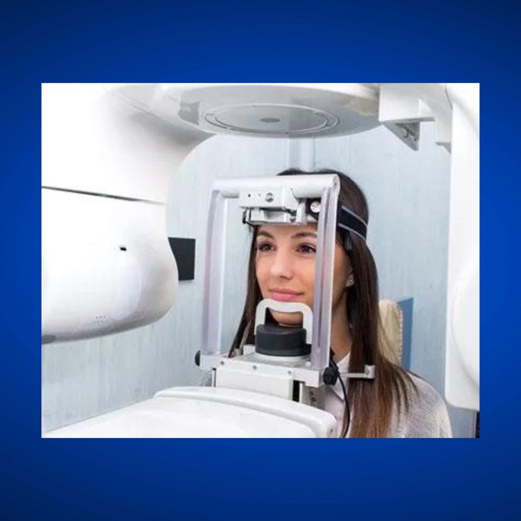
Unlike medical CT scanners that require large rooms and expose patients to significant radiation, dental Conebeam systems are compact, efficient, and use much lower radiation doses while providing superior image quality for oral and maxillofacial structures. The cone-shaped X-ray beam captures a complete 3D dataset in a single rotation around the patient’s head.
The resulting images can be viewed from any angle, sliced into cross-sections, and manipulated using sophisticated software to reveal information that would be impossible to obtain from conventional radiographs. This comprehensive visualization capability revolutionizes treatment planning and improves patient outcomes across all areas of dentistry.
Modern Conebeam systems provide sub-millimeter resolution, allowing Dr. Deaton to see fine anatomical details and detect problems in their earliest stages. This precision is particularly valuable for complex procedures requiring exact measurements and careful planning to ensure optimal results.
Advanced Diagnostic Capabilities
3D imaging reveals the exact position and orientation of impacted teeth, including wisdom teeth, canines, and other teeth that have failed to erupt properly. This detailed information allows for precise surgical planning that minimizes tissue trauma and reduces recovery time.
Root canal anatomy is clearly visualized in three dimensions, showing the number, location, and curvature of root canals that might be missed on traditional 2D radiographs. This comprehensive view helps ensure complete treatment and reduces the risk of missed canals that could lead to treatment failure.
Periodontal bone defects are accurately measured and characterized using 3D imaging, providing crucial information for treatment planning and prognosis assessment. The ability to see bone loss from multiple angles helps determine the most appropriate treatment approaches.
TMJ analysis benefits tremendously from 3D imaging, which shows the position and movement of jaw joints in detail that’s impossible with conventional X-rays. This information is invaluable for diagnosing and treating temporomandibular disorders.
Pathological lesions including cysts, tumors, and infections are clearly delineated in 3D images, showing their exact size, location, and relationship to adjacent structures. This detailed information is crucial for treatment planning and surgical approach selection.
Precise Implant Planning and Placement
Dental implant treatment planning has been revolutionized by 3D Conebeam imaging, which allows Dr. Deaton to evaluate bone quality, density, and volume with unprecedented precision. This detailed assessment ensures optimal implant placement and long-term success.
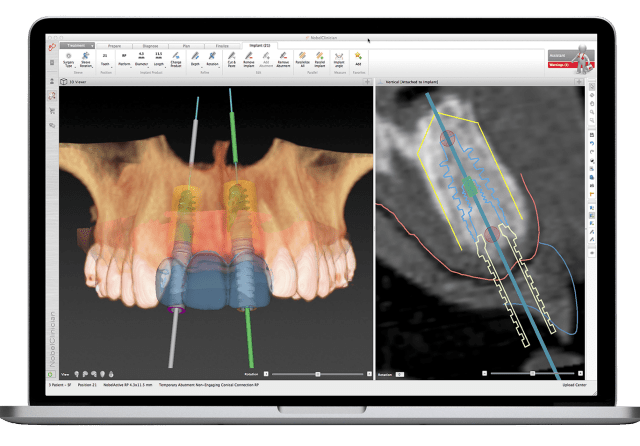
Virtual implant placement using specialized software allows Dr. Deaton to position implants precisely in the ideal location before surgery begins. This computer-guided planning improves accuracy while reducing surgical time and patient discomfort.
Bone density measurements obtained from 3D scans help determine the most appropriate implant design and surgical protocol for each patient. This personalized approach optimizes success rates and reduces the risk of implant failure.
Anatomical structure identification including nerves, sinuses, and blood vessels ensures safe implant placement without damage to vital structures. The ability to see these structures clearly in 3D prevents complications and improves surgical outcomes.
Guided surgery protocols use 3D treatment plans to create surgical guides that transfer the virtual plan to the actual surgery. This technology ensures that implants are placed exactly as planned with minimal deviation.
Comprehensive Orthodontic Analysis
Orthodontic treatment planning benefits significantly from 3D imaging, which provides detailed information about tooth positions, root orientations, and bone support that’s crucial for successful tooth movement. This technology helps predict treatment outcomes and identify potential complications.
Airway analysis using 3D imaging can identify breathing problems related to oral and facial structure that may benefit from orthodontic treatment. This information helps create treatment plans that improve both aesthetics and function.
Root resorption assessment is more accurate with 3D imaging, allowing orthodontists to monitor for this potential complication and modify treatment as needed to prevent damage to tooth roots.
Impacted tooth evaluation provides detailed information about the position and orientation of teeth that have failed to erupt, helping determine whether orthodontic movement is possible or if surgical exposure is necessary.
Treatment progress monitoring using periodic 3D scans allows for precise assessment of tooth movement and treatment effectiveness, enabling adjustments to optimize outcomes.
Advanced Oral Surgery Applications
Complex extractions benefit from 3D planning, which shows the exact position of tooth roots relative to nerves, sinuses, and adjacent teeth. This information allows for safer, more efficient removal with reduced risk of complications.
Bone grafting procedures are planned more precisely using 3D imaging to assess existing bone volume and plan grafting approaches that optimize outcomes. The technology helps determine the amount and type of graft material needed.
Sinus lift procedures require detailed knowledge of sinus anatomy that 3D imaging provides. The technology shows sinus size, shape, and the presence of septa or other anatomical variations that affect surgical planning.
Facial trauma assessment uses 3D imaging to evaluate fractures and plan reconstructive procedures. The detailed visualization helps surgeons understand the extent of damage and plan appropriate treatment approaches.
Pre-surgical planning using 3D images allows surgeons to practice complex procedures virtually before performing them on patients. This preparation improves efficiency and reduces surgical time and complications.
Endodontic Treatment Enhancement
Root canal treatment planning is significantly improved with 3D imaging, which reveals root canal anatomy that may not be visible on traditional radiographs. This comprehensive view helps ensure complete treatment and reduces failure rates.
Missed canal detection is enhanced with 3D imaging, which can reveal additional canals that might be overlooked during conventional treatment. Finding and treating all canals is crucial for endodontic success.
Retreatment evaluation benefits from 3D imaging, which shows the quality of previous root canal treatment and helps identify reasons for failure. This information guides retreatment planning and improves success rates.
Surgical endodontics planning uses 3D imaging to plan apicoectomy procedures and identify the exact location of root tips relative to anatomical structures. This precision improves surgical outcomes and reduces complications.
Perforation assessment and repair planning benefits from the detailed visualization that 3D imaging provides, helping endodontists understand the extent of perforations and plan appropriate repair procedures.
Periodontal Disease Assessment
Bone loss measurement is more accurate with 3D imaging, which shows the exact extent and pattern of periodontal bone destruction. This information helps determine prognosis and plan appropriate treatment approaches.
Furcation involvement in multi-rooted teeth is clearly visualized with 3D imaging, showing the degree of bone loss between roots that’s difficult to assess with conventional radiographs.
Regenerative therapy planning benefits from detailed 3D assessment of bone defects, helping periodontists select appropriate materials and techniques for optimal regeneration outcomes.
Implant site assessment after periodontal treatment uses 3D imaging to evaluate bone healing and determine the optimal timing and placement of dental implants.
Treatment monitoring using periodic 3D scans allows for precise assessment of healing and treatment effectiveness, enabling modifications to optimize outcomes.
Radiation Safety and Dose Optimization
Modern Conebeam systems use significantly less radiation than medical CT scanners while providing superior image quality for dental applications. Advanced dose reduction technologies minimize patient exposure while maintaining diagnostic quality.
Collimation techniques focus the X-ray beam only on the area of interest, reducing unnecessary radiation exposure to surrounding tissues. This precision targeting optimizes the risk-benefit ratio of 3D imaging.
Automatic exposure controls adjust radiation dose based on patient size and density, ensuring optimal image quality with minimal radiation exposure. These systems prevent overexposure while maintaining diagnostic capability.
Protocol optimization for different clinical applications ensures that patients receive only the radiation necessary for their specific diagnostic needs. Customized protocols balance image quality with dose reduction.
Regular calibration and quality assurance procedures ensure that Conebeam systems operate at peak efficiency with minimal radiation output. This attention to equipment maintenance protects patient safety.
Image Analysis and Interpretation
Specialized software allows for comprehensive analysis of 3D images, including measurements, virtual surgery planning, and simulation of treatment outcomes. These tools enhance diagnostic accuracy and treatment precision.
Multi-planar reconstruction creates views in any desired plane, allowing Dr. Deaton to examine structures from optimal angles for diagnosis and treatment planning. This flexibility provides comprehensive visualization.
Volume rendering techniques create realistic 3D images that help patients understand their anatomy and treatment needs. These visualizations improve patient education and treatment acceptance.
Quantitative analysis tools measure bone density, volume, and other parameters crucial for treatment planning. These objective measurements support evidence-based treatment decisions.
Comparison capabilities allow current images to be compared with previous scans to monitor changes over time. This temporal analysis helps assess treatment progress and disease progression.
Patient Education and Communication
3D visualizations help patients understand their oral health conditions and treatment needs better than ever before. Seeing their own anatomy in three dimensions makes complex problems and treatments easier to comprehend.
Virtual treatment simulations show patients what to expect from proposed procedures, improving informed consent and reducing anxiety about treatment. These demonstrations enhance communication and treatment acceptance.
Interactive software allows Dr. Deaton to manipulate 3D images during patient consultations, pointing out specific problems and explaining treatment approaches clearly and effectively.
Printed 3D images can be provided to patients who want to share their diagnostic information with other healthcare providers or keep personal copies for their records.
Before-and-after comparisons using 3D imaging demonstrate treatment outcomes clearly, helping patients understand the value of completed treatment and motivating continued care.
Integration with Treatment Planning
CAD/CAM integration allows 3D images to be used directly in the design and manufacture of dental restorations, ensuring optimal fit and function based on precise anatomical data.
Surgical guide fabrication uses 3D image data to create precise guides that transfer treatment plans from computer planning to actual procedures. This technology improves accuracy and reduces surgical time.
Prosthetic planning benefits from detailed 3D anatomy information, allowing for optimal design of crowns, bridges, and dentures that fit precisely and function optimally.
Orthodontic appliance design uses 3D data to create custom brackets, wires, and aligners that provide optimal tooth movement with maximum efficiency and comfort.
Treatment simulation allows Dr. Deaton to test different treatment approaches virtually before beginning actual treatment, optimizing outcomes and reducing the need for treatment modifications.
Special Applications and Advanced Techniques
Sleep apnea evaluation uses 3D imaging to assess airway dimensions and identify anatomical factors contributing to breathing problems. This information helps guide treatment selection and planning.
Facial asymmetry analysis benefits from 3D imaging, which provides precise measurements and visualizations of facial structures that help plan corrective procedures.
Growth and development monitoring in children uses 3D imaging to track changes over time and identify problems that may require early intervention.
Cleft palate evaluation and treatment planning uses detailed 3D visualization to understand complex anatomical relationships and plan comprehensive treatment approaches.
Research applications of 3D imaging contribute to advancing dental knowledge and improving treatment techniques through detailed anatomical studies and outcome assessments.
Quality Assurance and Maintenance
Regular calibration ensures that 3D imaging equipment maintains optimal performance and produces accurate, consistent images. This attention to quality control protects diagnostic accuracy.
Staff training programs ensure that all team members understand proper operation of 3D imaging equipment and can provide patients with accurate information about the technology and procedures.
Image quality monitoring systems track performance parameters and identify potential problems before they affect diagnostic capability or patient care.
Equipment maintenance protocols ensure continuous operation and prevent equipment failures that could disrupt patient care or compromise image quality.
Backup and storage systems protect 3D image data against loss while ensuring accessibility for future reference and comparison studies.
Cost-Effectiveness and Value
While 3D imaging represents an investment in advanced technology, the improved diagnostic accuracy and treatment precision often result in better outcomes and fewer complications, providing excellent long-term value.
Reduced treatment time and fewer complications translate to cost savings for patients through more efficient procedures and reduced need for retreatment or additional procedures.
Enhanced treatment planning reduces the likelihood of surgical complications that could require additional treatment or extended recovery periods.
Improved treatment outcomes and patient satisfaction justify the investment in advanced 3D imaging technology through better results and enhanced patient experiences.
Insurance coverage for 3D imaging is increasingly available when the technology is medically necessary for diagnosis or treatment planning, making advanced imaging more accessible to patients.
Why Choose Adamsville Family Dentistry for 3D Imaging
Dr. Deaton’s investment in state-of-the-art 3D Conebeam imaging technology demonstrates our commitment to providing the most advanced diagnostic capabilities available in dentistry today. This technology enables more accurate diagnoses and precise treatment planning.
Our comprehensive approach to 3D imaging includes proper training, quality assurance, and patient education to ensure that you receive the full benefits of this revolutionary technology. We believe that advanced diagnostics lead to better treatment outcomes.
Located in Adamsville, our practice has earned a reputation for utilizing cutting-edge dental technology to provide superior patient care. We understand that precise diagnosis and planning are essential for optimal treatment results.
The integration of 3D imaging with our comprehensive dental services allows for seamless treatment planning and execution, providing patients with the most advanced care available in a comfortable, familiar environment.
Experience the Future of Dental Diagnostics
Don’t settle for limited diagnostic information from traditional 2D X-rays when advanced 3D imaging can provide comprehensive visualization of your oral structures. This technology enables more accurate diagnoses and precise treatment planning for optimal outcomes.
The detailed information provided by 3D Conebeam imaging allows Dr. Deaton to plan and perform procedures with unprecedented precision, improving success rates while reducing complications and recovery time.
Whether you need implant placement, complex extraction, orthodontic treatment, or oral surgery, 3D imaging provides the detailed information necessary for optimal treatment planning and execution.
Discover the difference that advanced 3D imaging can make in your dental care. Call Adamsville Family Dentistry at 731-632-3371 to learn more about our 3D Conebeam imaging capabilities and how this technology can improve your treatment outcomes, or schedule your appointment now to experience the most advanced diagnostic imaging available in dentistry and benefit from the precision, safety, and superior results that 3D technology provides.
Routine Check-Ups
Regular dental visits help detect issues early, ensuring optimal oral health and preventing major problems.
Professional Cleanings
Our thorough cleanings remove plaque and tartar buildup, reducing the risk of cavities and gum disease.
Tooth-Colored Fillings
We use high-quality, tooth-colored fillings to restore damaged teeth and protect them from further decay.
Tooth Extractions
When necessary, we perform extractions safely and comfortably to maintain overall dental health.